• Home
  • Huvitz Câmera de Fundo de Olho HFC-1

Huvitz Câmera de Fundo de Olho HFC-1

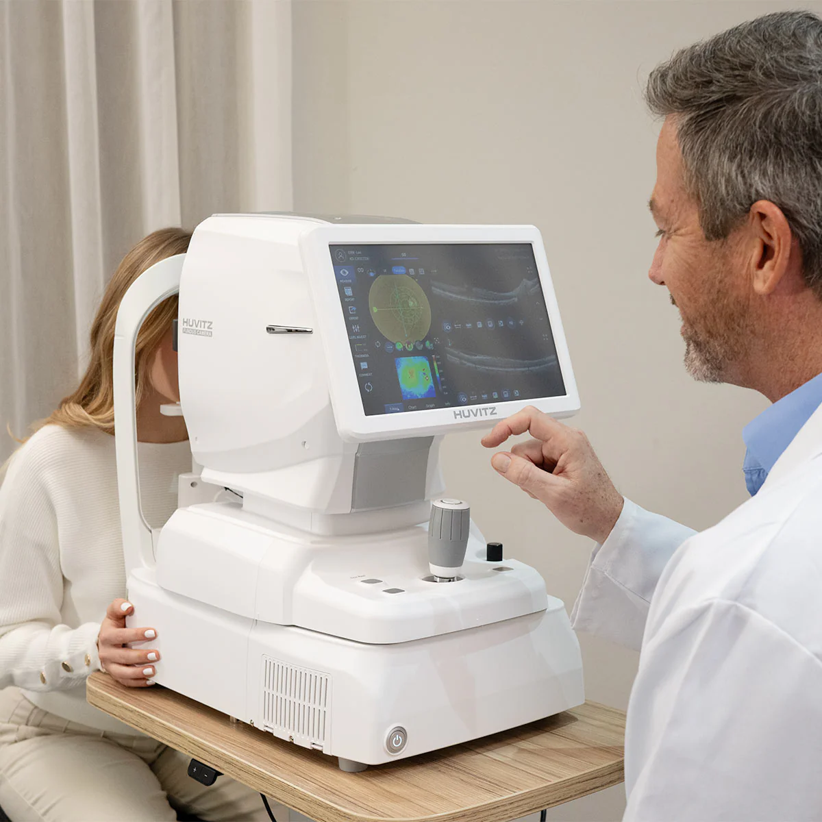
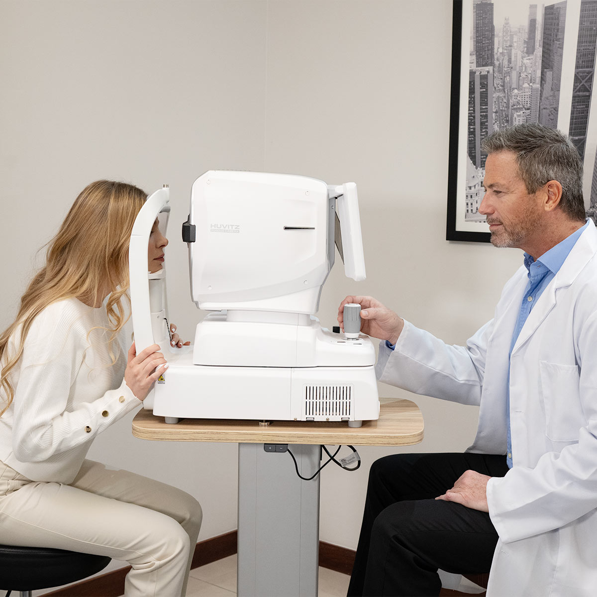
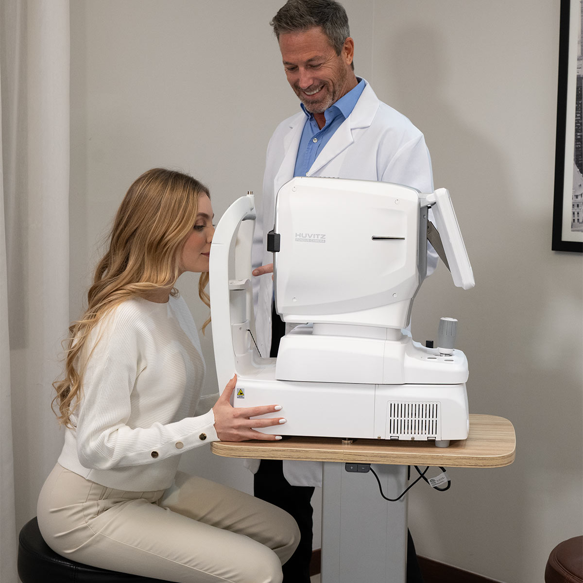
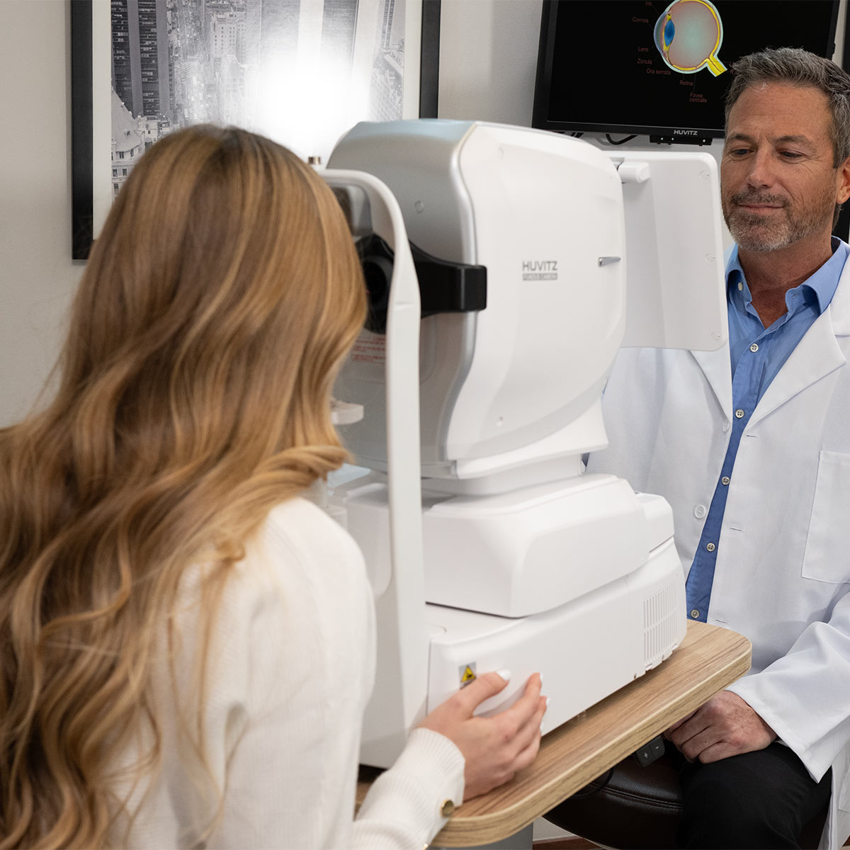
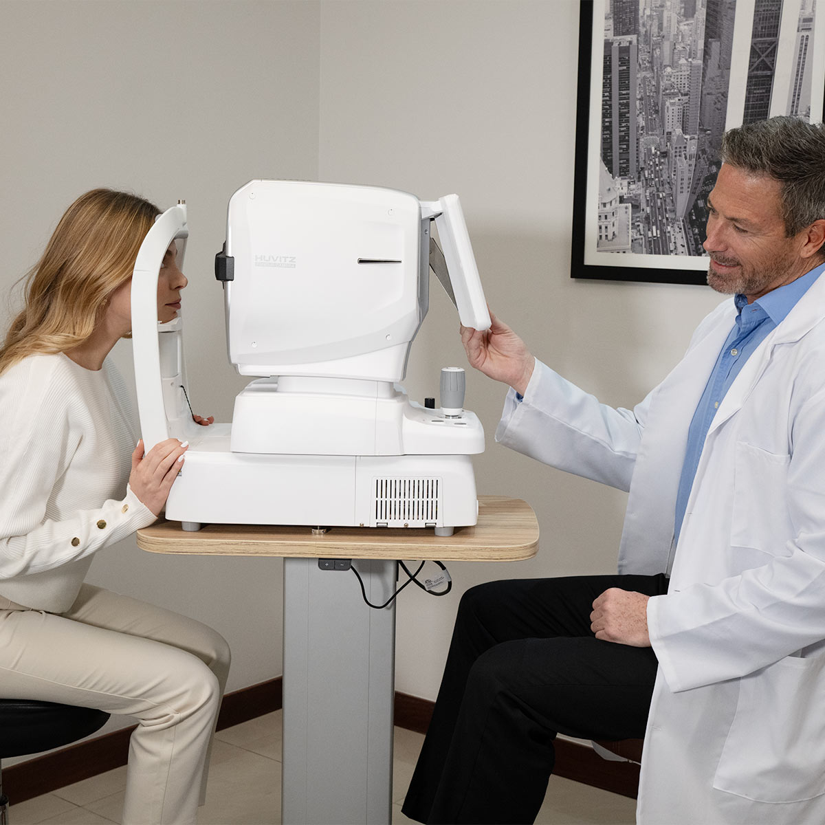
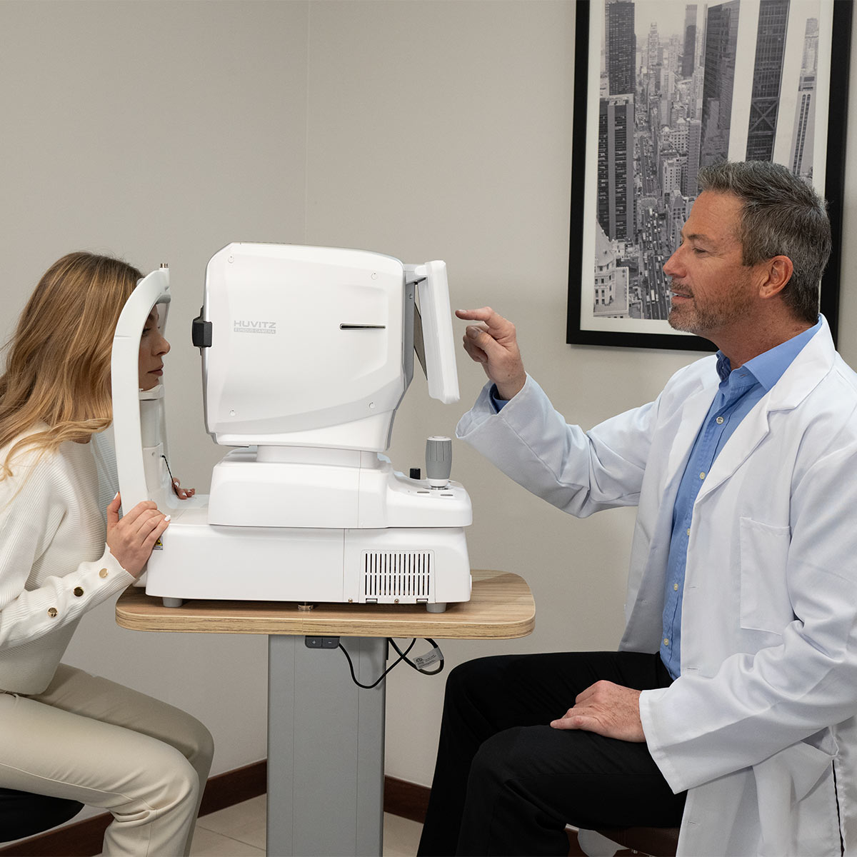
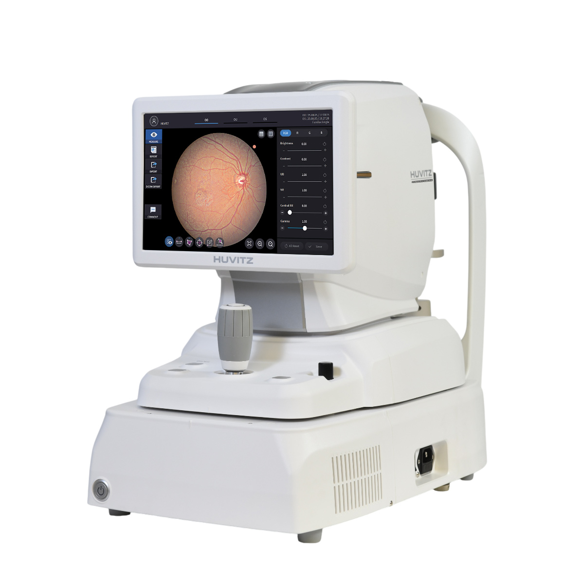
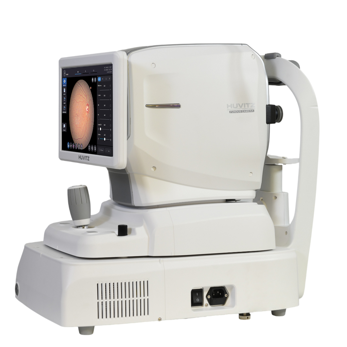
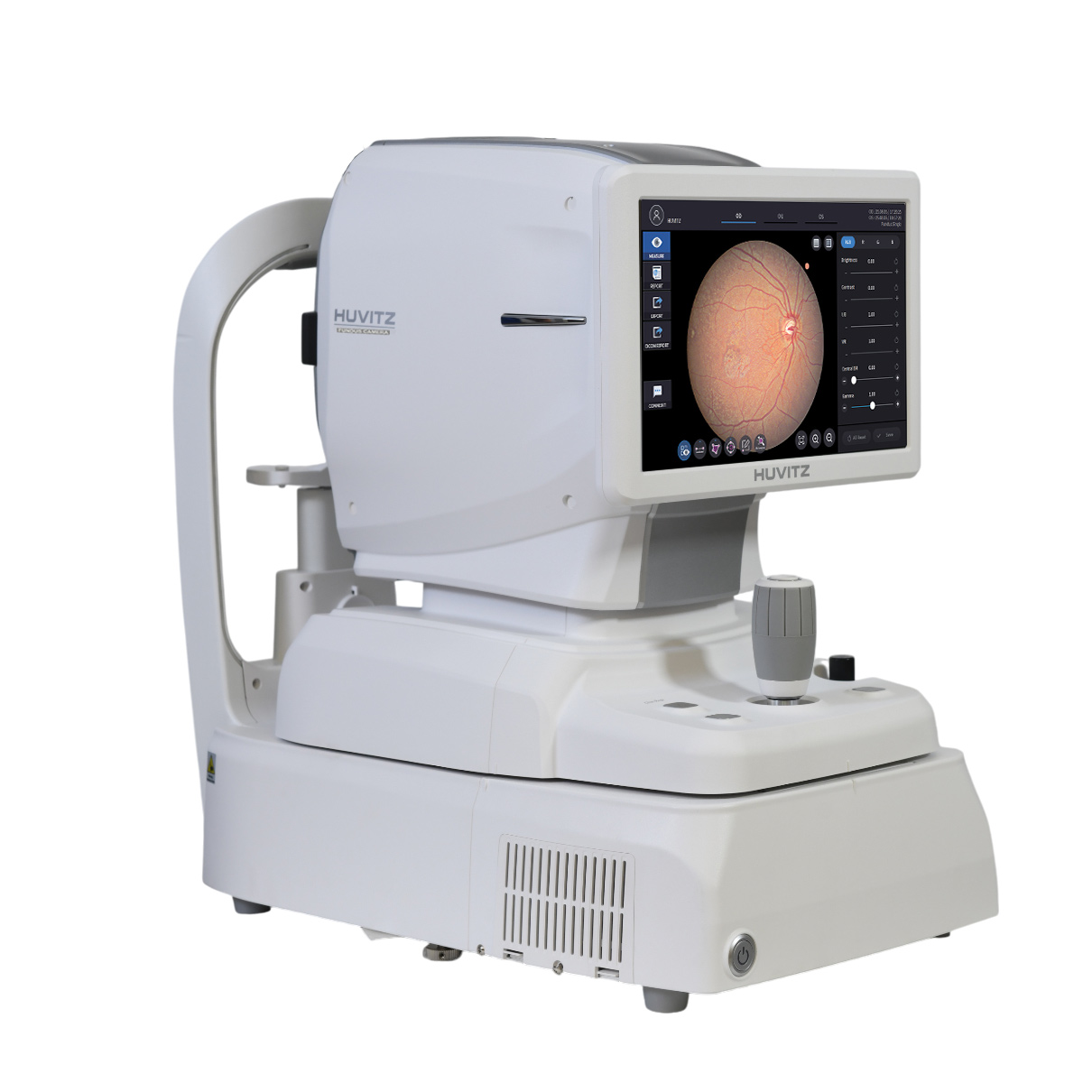
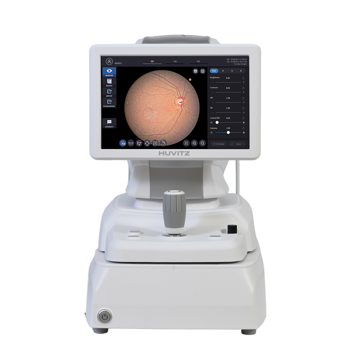
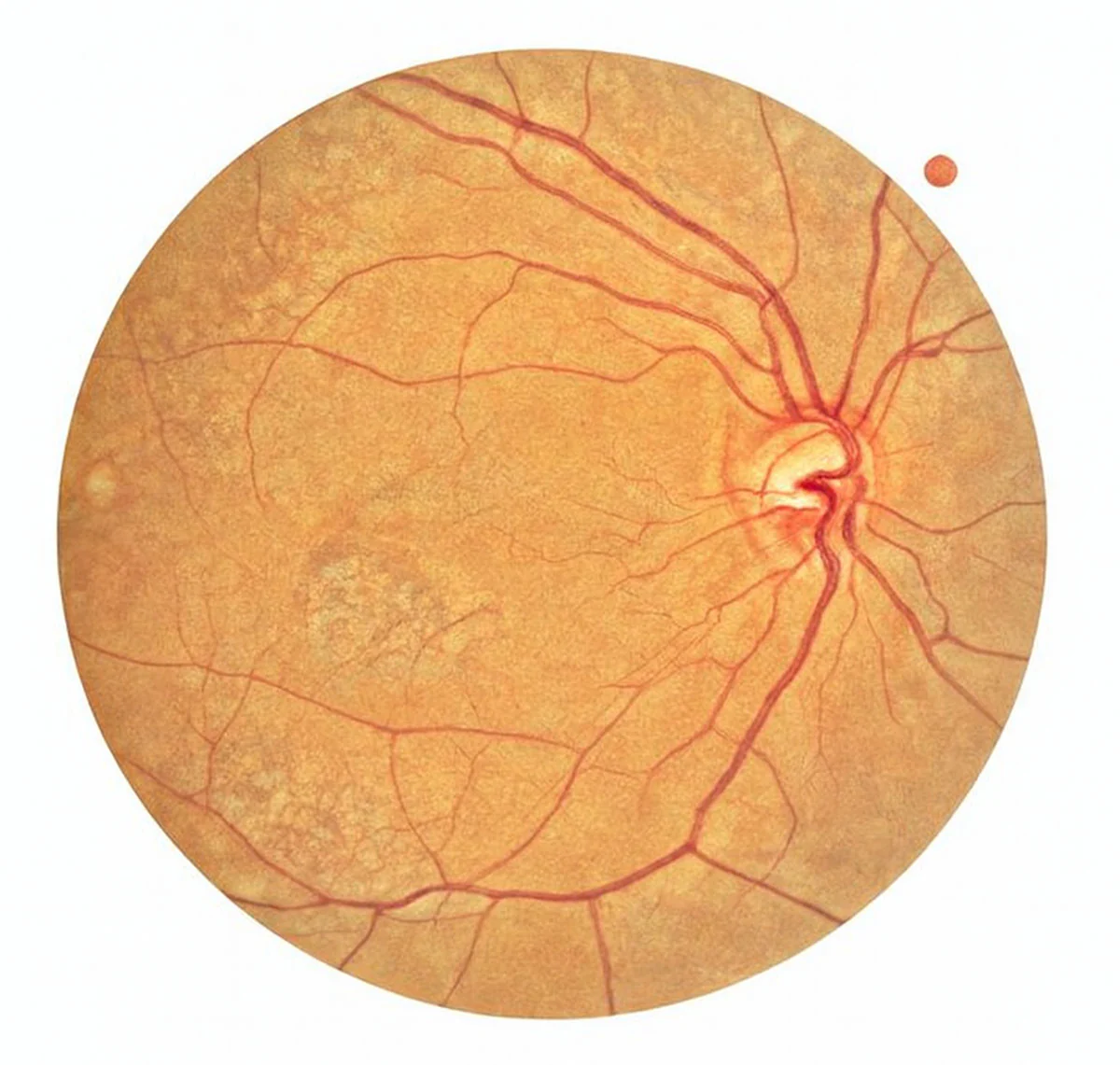
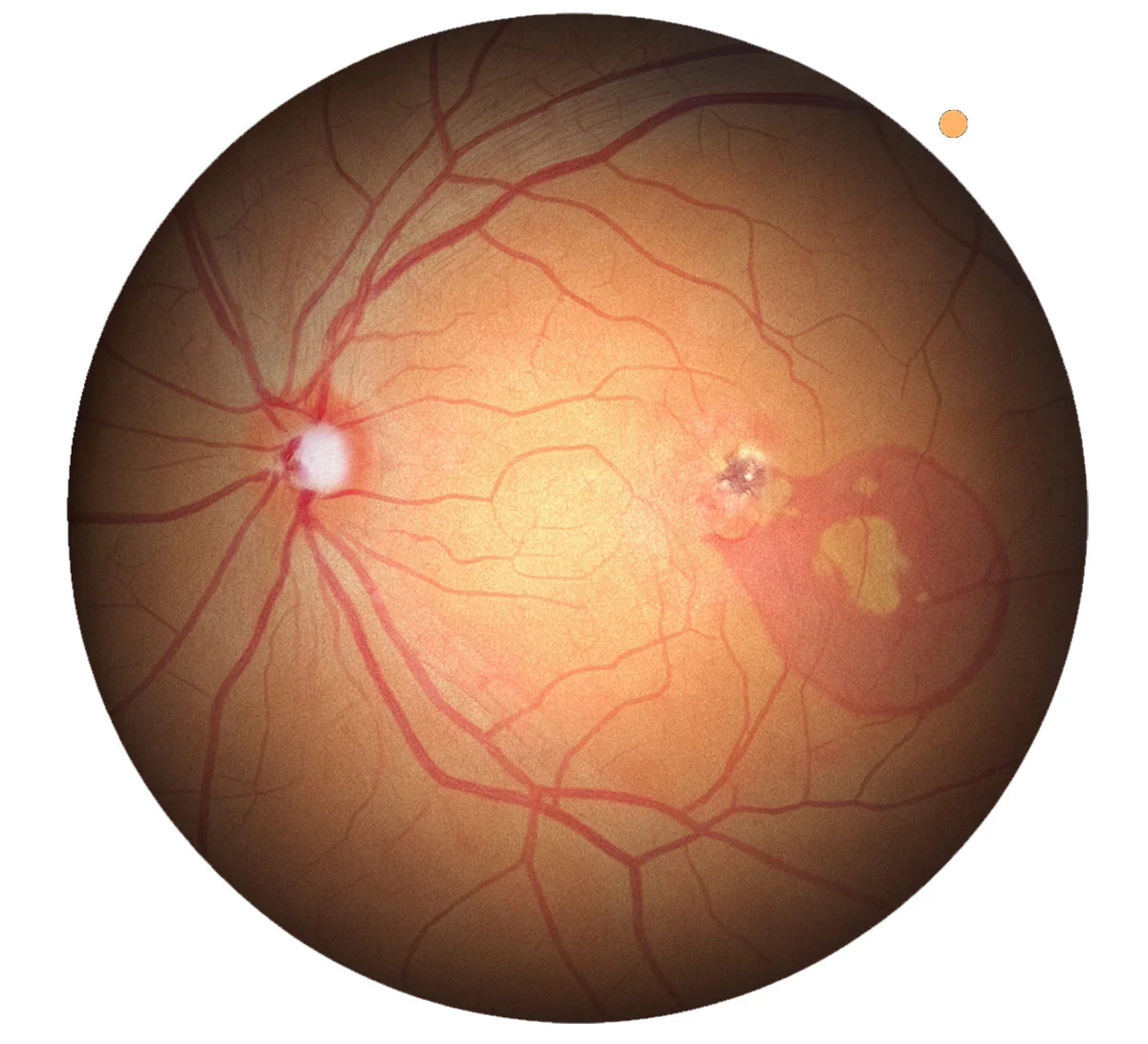
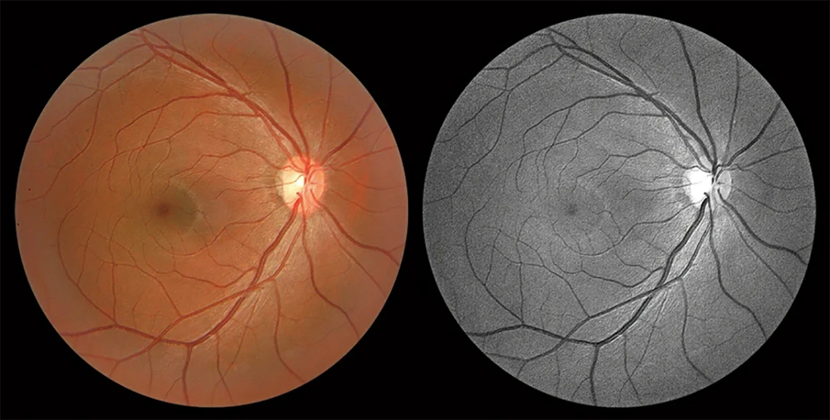
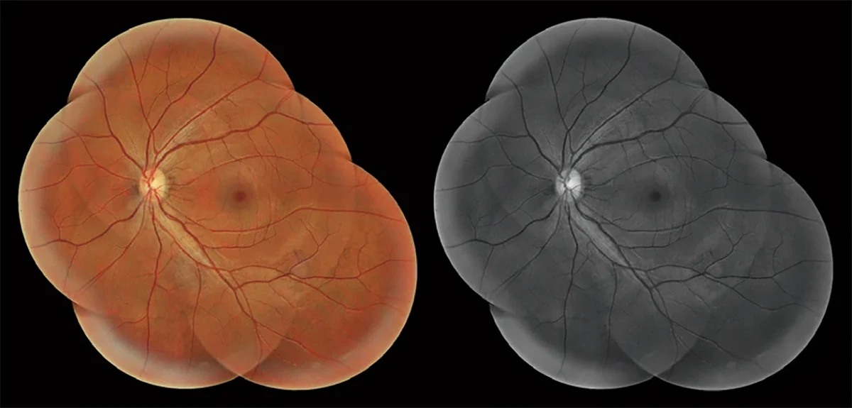
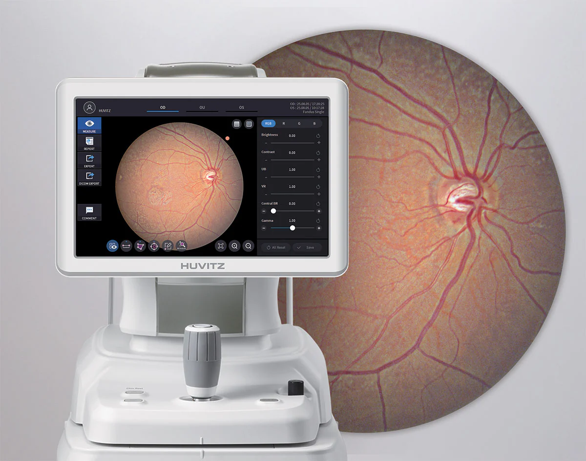
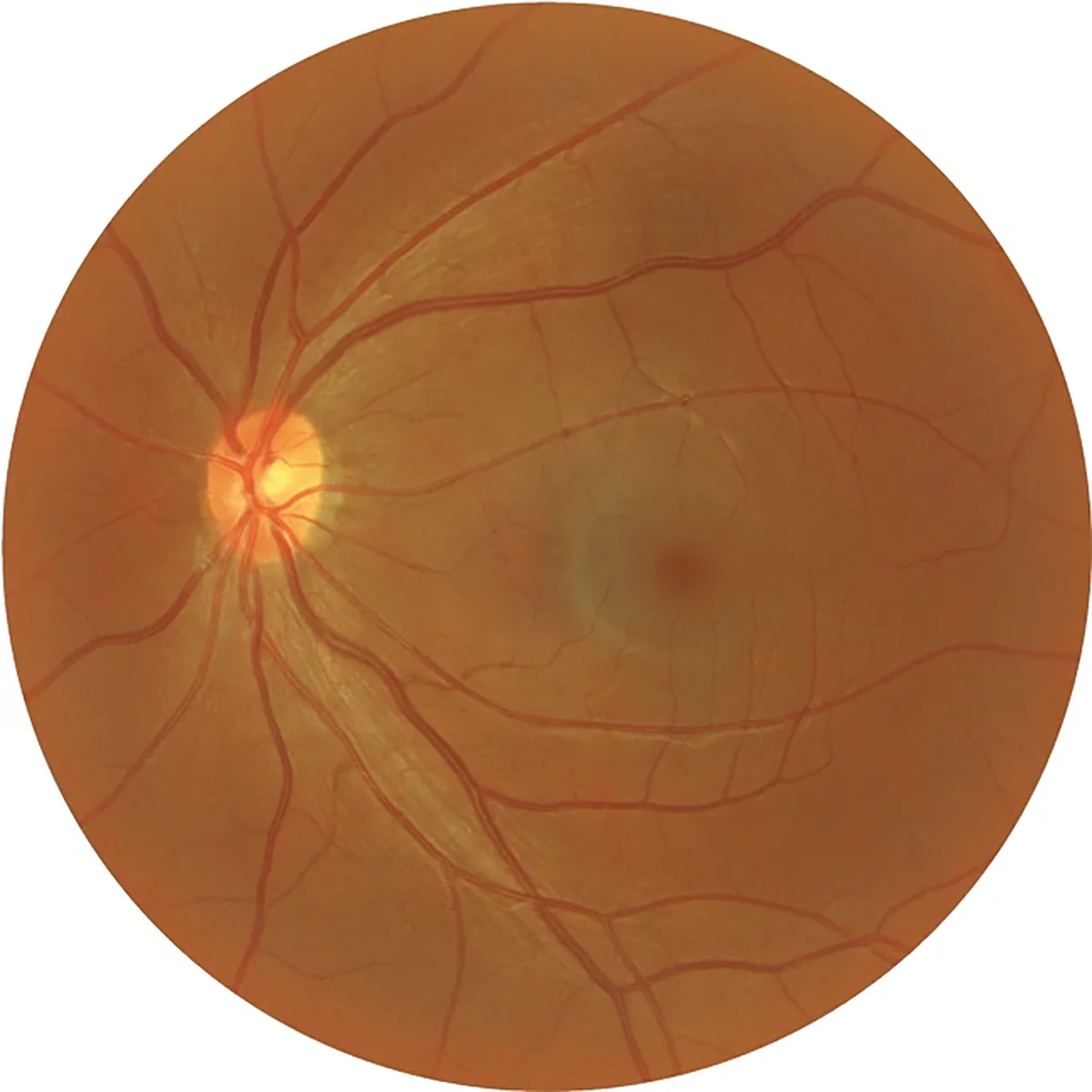
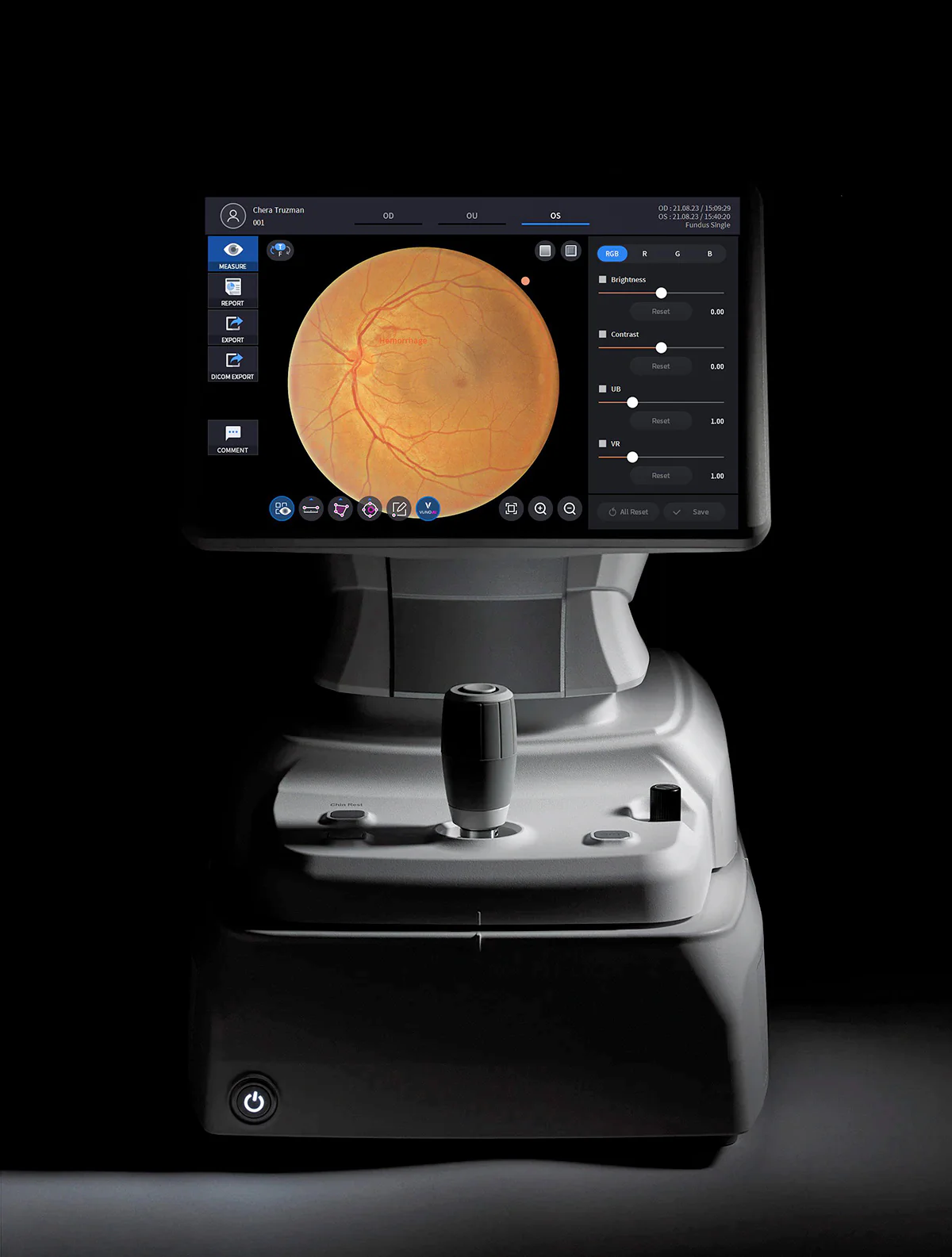
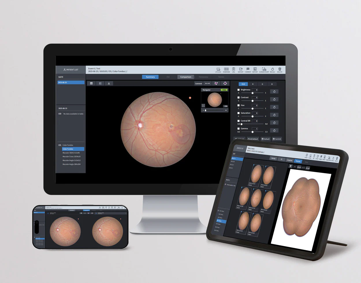
O rastreamento e disparo automáticos permitem que a Câmera de Fundo HFC-1 ajuste os modos de forma rápida e estável por conta própria, enquanto mede diferentes tamanhos de pupila. Sua câmera de alta definição de 20 megapixels captura imagens com menor artefato de movimento e possui a capacidade de ampliar as imagens para estudar detalhes finos. A nova Câmera de Fundo HFC-1 inclui uma variedade de modos de imagem: colorido, azul, vermelho, sem vermelho e cobalto, que auxiliam nos exames de Glaucoma, RNFL, Edema, Anomalias Pigmentares e mais. Os profissionais de saúde ocular podem medir, analisar, diagnosticar e gerar relatórios no próprio local utilizando o PC integrado da Câmera de Fundo, com sua prática tela sensível ao toque LCD de 12,1” para navegação.

Falar com um especialista

Imagens de Retina em Alta Definição

Captura imagens nítidas e detalhadas do fundo de olho, permitindo diagnósticos mais precisos e confiáveis.

Captura Rápida e Operação Simplificada

Sistema otimizado que permite aquisição rápida de imagens, reduzindo o tempo de exame e aumentando a eficiência clínica.

Interface Intuitiva e Controle Inteligente

Tela moderna e navegação simples facilitam a operação diária, garantindo maior produtividade e facilidade de uso.

Integração Digital e Compartilhamento Fácil

Compatível com sistemas digitais, permitindo armazenar, analisar e compartilhar imagens com rapidez e segurança.

User-Selected Fundus Color Mode:
Four fundus image color tone options are available, allowing the user to choose the color mode best suited to their diagnostic style or visual preference. This provides a more accurate and comfortable observation of the retinal blood vessels.

Improved Lesion Visibility with Adaptive Brightness and Color Control:
By adjusting the Central BR and Gamma values, it is possible to refine the brightness and color balance of the fundus image, allowing for clearer visualization of the areas where lesions are located. The HFC-1’s Advanced Visualization Technology (EVT) ensures the capture of high-quality images in various clinical scenarios, being especially effective in identifying subtle pathological changes.

Target Fixation:
By adjusting the position of the Target Fixation on the screen, users can obtain even more precise measurement results.

Panorama Function:
This function provides essential information for the overall assessment of the eyes, allowing the capture of high-resolution images with minimal distortion.

True-Color Fundus Scan in a Single Shot:
With 12-bit color depth and advanced gamma correction, the system captures true-to-color fundus images in a single shot—completely free of chromatic aberration. It offers uniform contrast between dark retinal regions and bright optical discs, allowing for clear visualization of arteries, veins, and fine microvasculature. Furthermore, it automatically aligns and combines up to seven images to create a wide-coverage panoramic view, facilitating intuitive identification of lesion location and extent in a single comprehensive image.

High-Performance 20 Megapixel Camera:
The efficient camera reduces motion artifacts and captures high-quality images, allowing for analysis from overview to fine detail magnification. Furthermore, images can be viewed in various modes, aiding in analysis and diagnosis.

Multiple Image Modes
: • Color : Bright, full-spectrum images
• Blue : RNFL, wrinkles, edema, cell membranes
• Red : Pigmentary changes, choroidal ruptures, nevi, melanoma
• Red-Free : Glaucoma, diabetic retinopathy
• Cobalt : RNFL

Compact Design for Space Savings
With a compact design and integrated 1TB PC, measurements, analyses, diagnostics, and reports can be performed in a single location. The HFC-1 was designed to optimize space usage.

12.1″ Touch Screen LCD
The HFC-1 offers high-resolution images without ghosting thanks to its real-time image processor. With the adoption of Wide Color TFT LCD , users view live images with excellent quality. Furthermore, the touchscreen significantly increases operational convenience.

Accurate Comparative Analysis Based on Detailed Information
Through comprehensive and detailed analyses, professionals can clearly understand the specific symptoms, conditions, and progression of each patient. The system allows for precise comparison of pre- and post-treatment images and supports assessments from multiple perspectives through Single and Panorama reports, offering a deeper understanding of pathological changes. Without the need to install additional software, users can view and analyze data directly in browsers such as Internet Explorer, Safari, and Chrome. With full DICOM compatibility, the HFC-1 keeps pace with the most advanced trends in medical IT integration.

Comprehensive Comparative Analysis for Advanced Diagnostics
: With complete and in-depth analyses, professionals can clearly understand the individual symptoms, conditions, and evolution of each patient. The system enables precise comparisons between pre- and post-treatment images and allows for multi-angle assessments of pathological changes through Single and Panorama reports.

Manuais e catalogos

Catalogos

HFC-1

Manuais

HFC-1