-
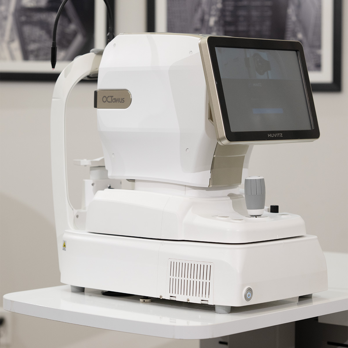
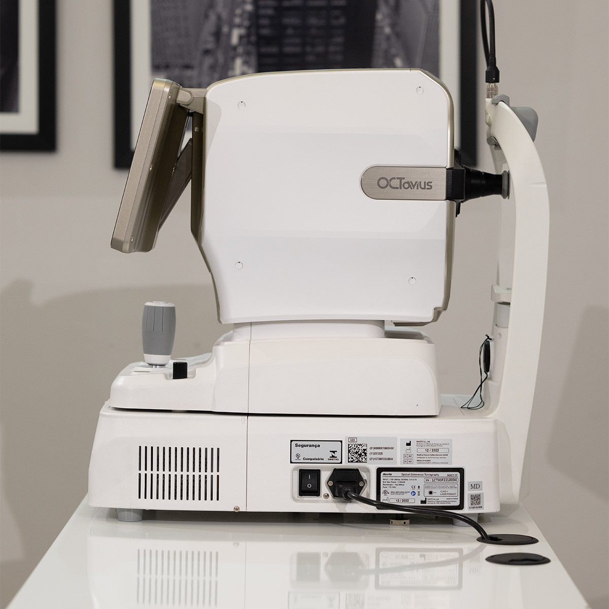
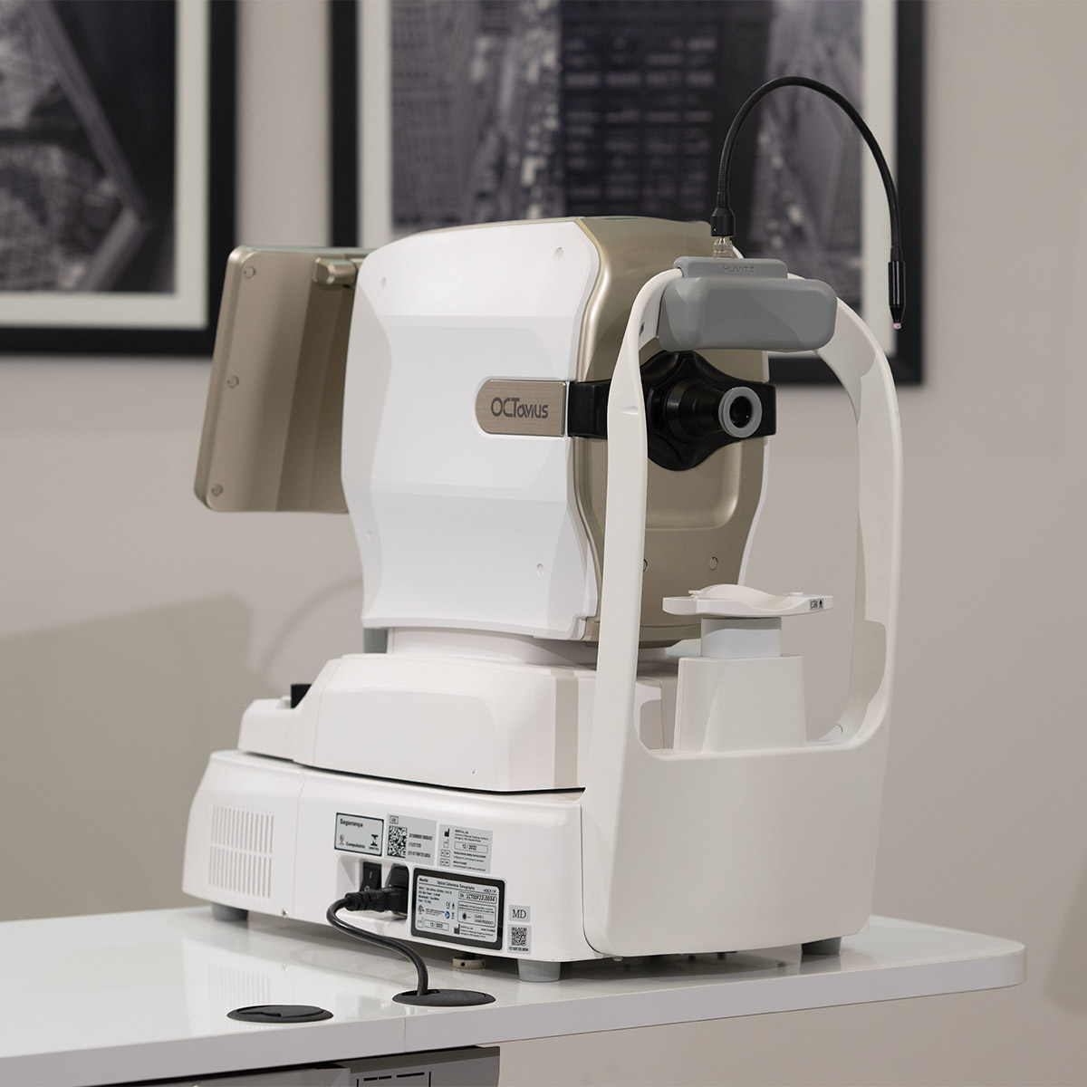
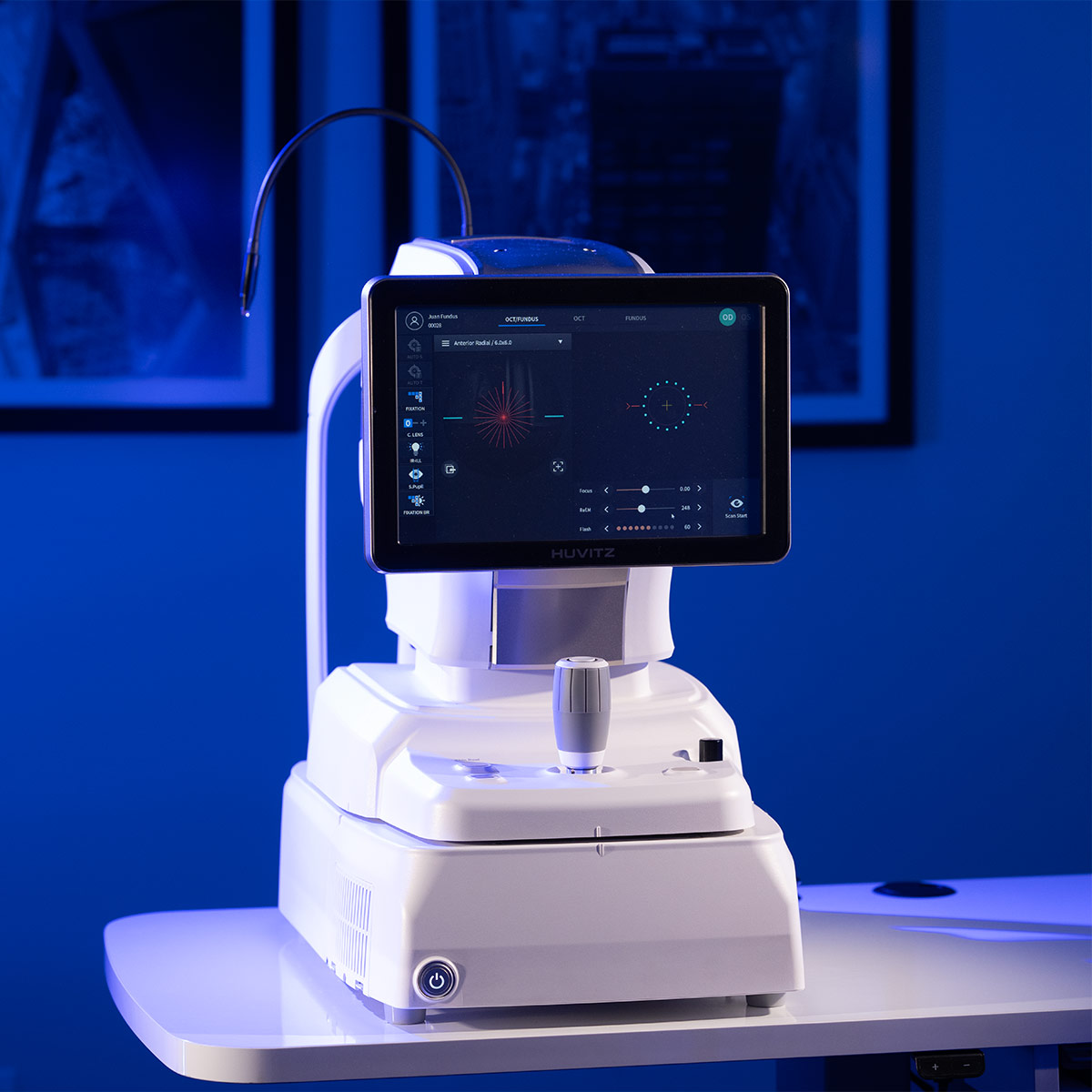
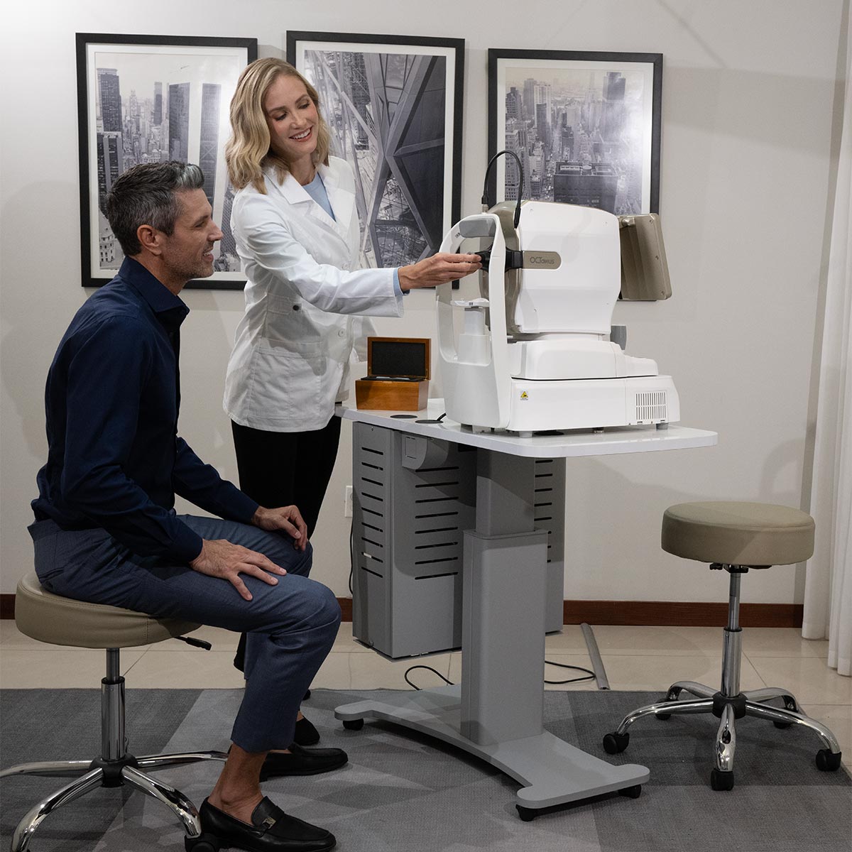
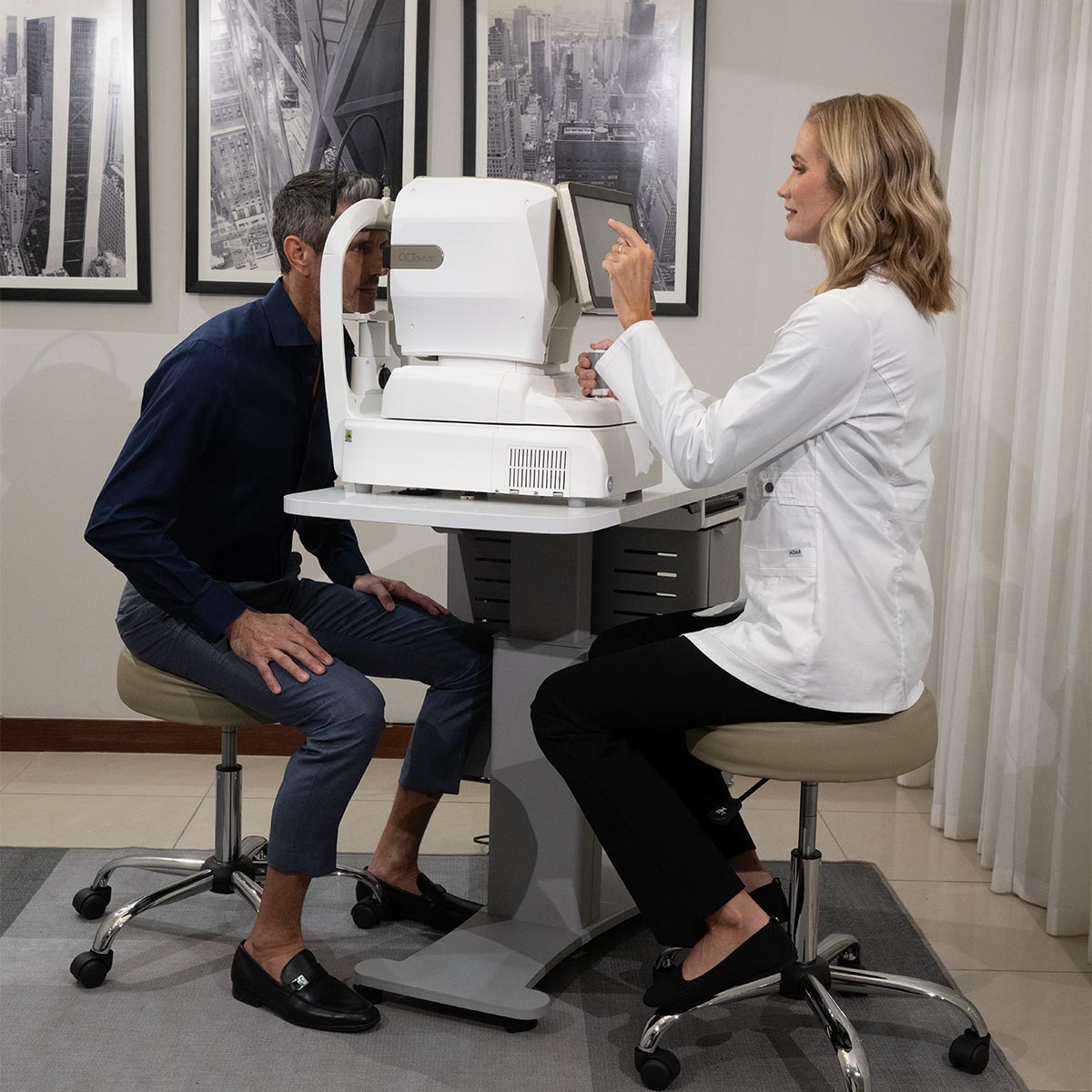
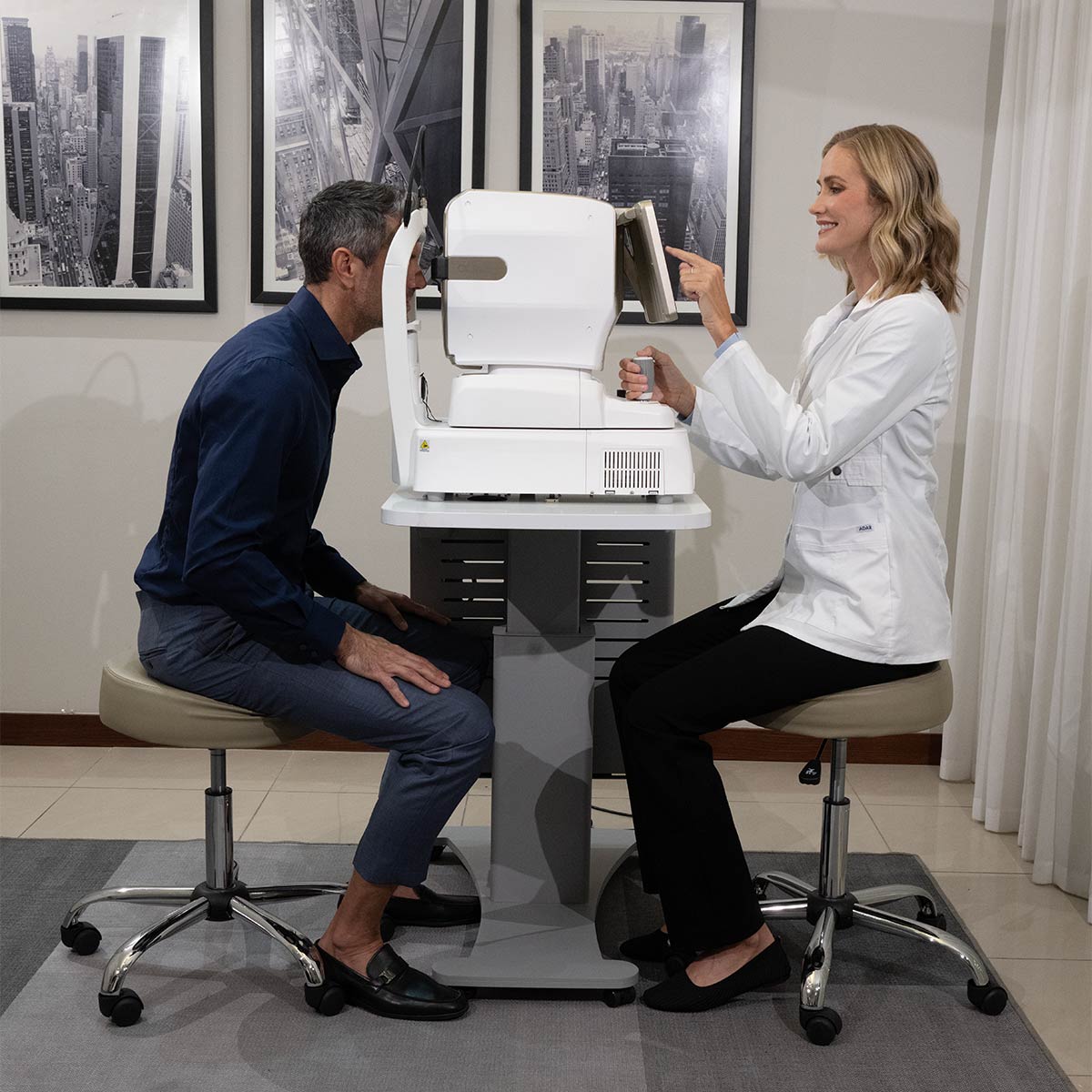
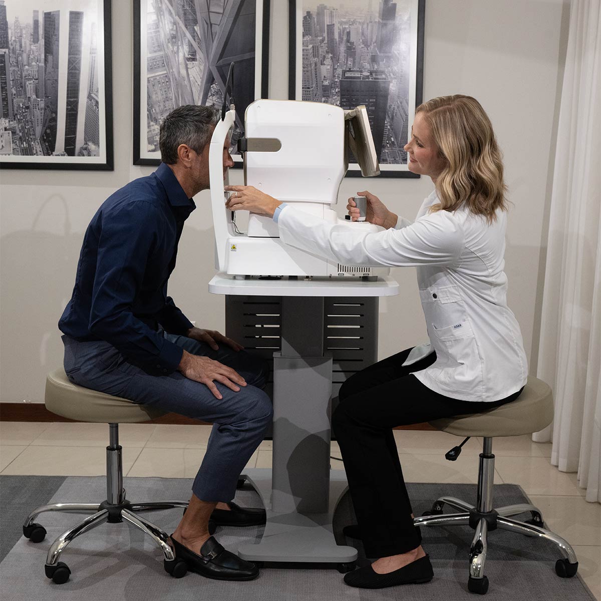
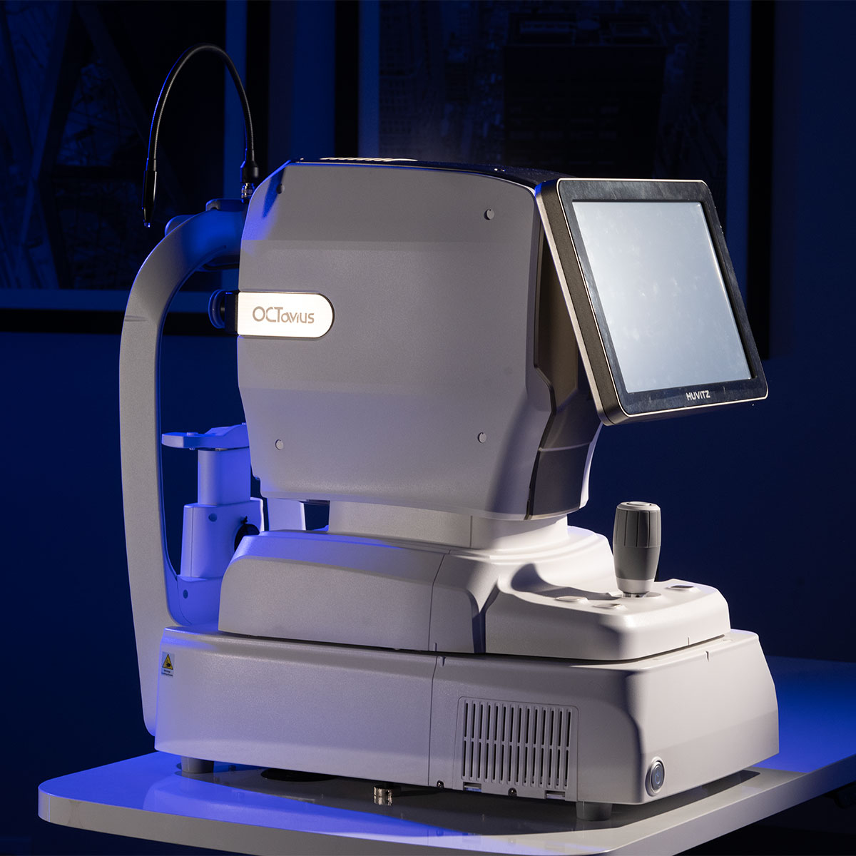
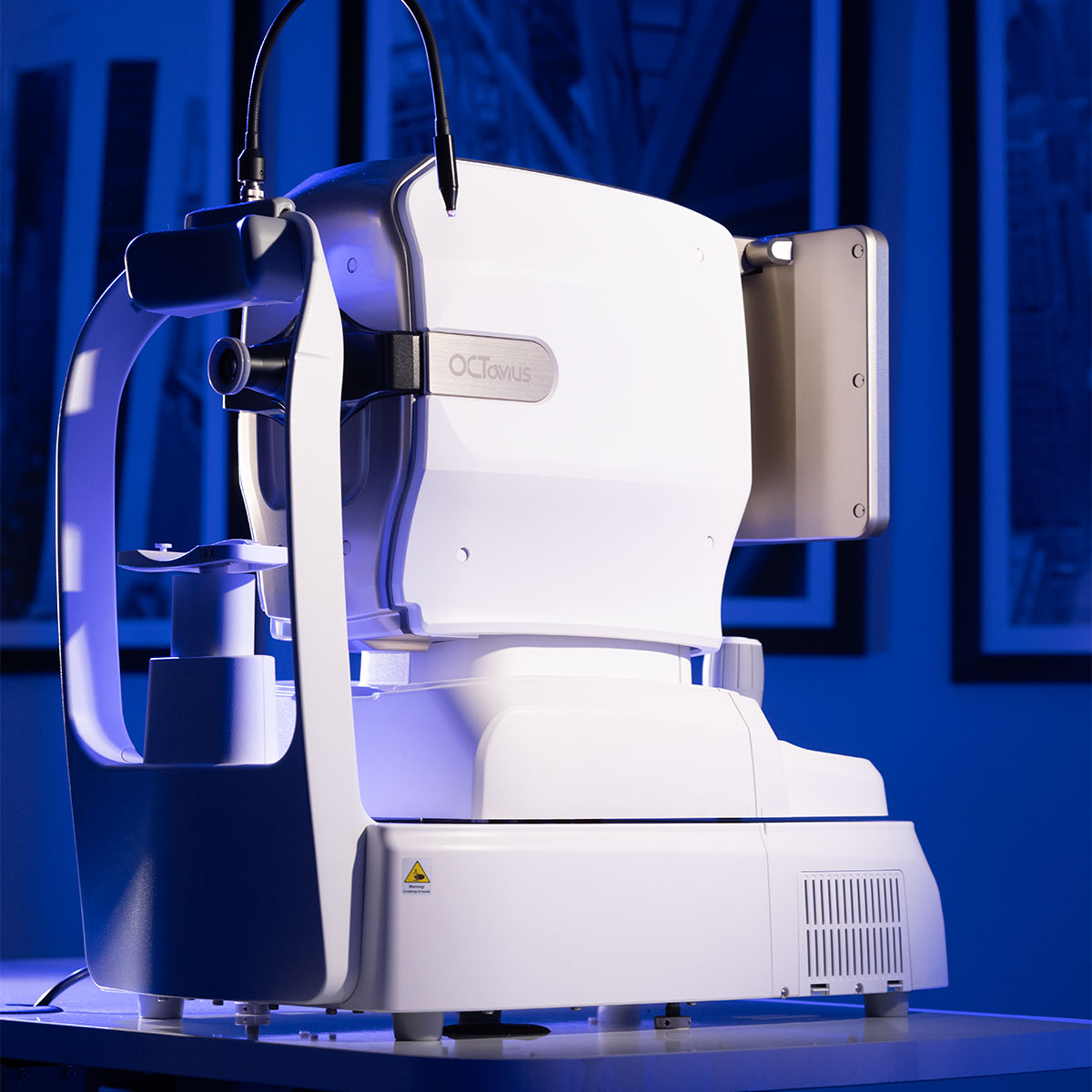
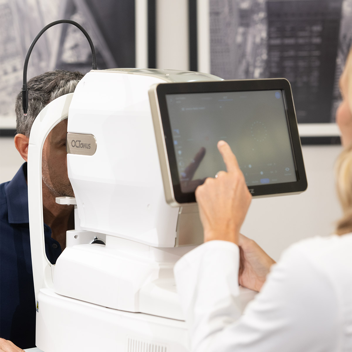
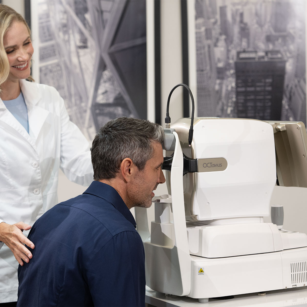
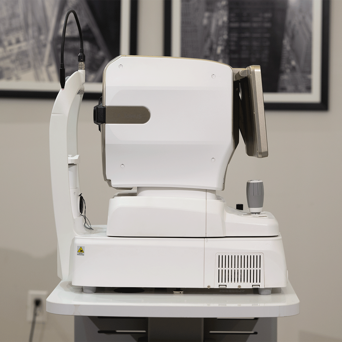
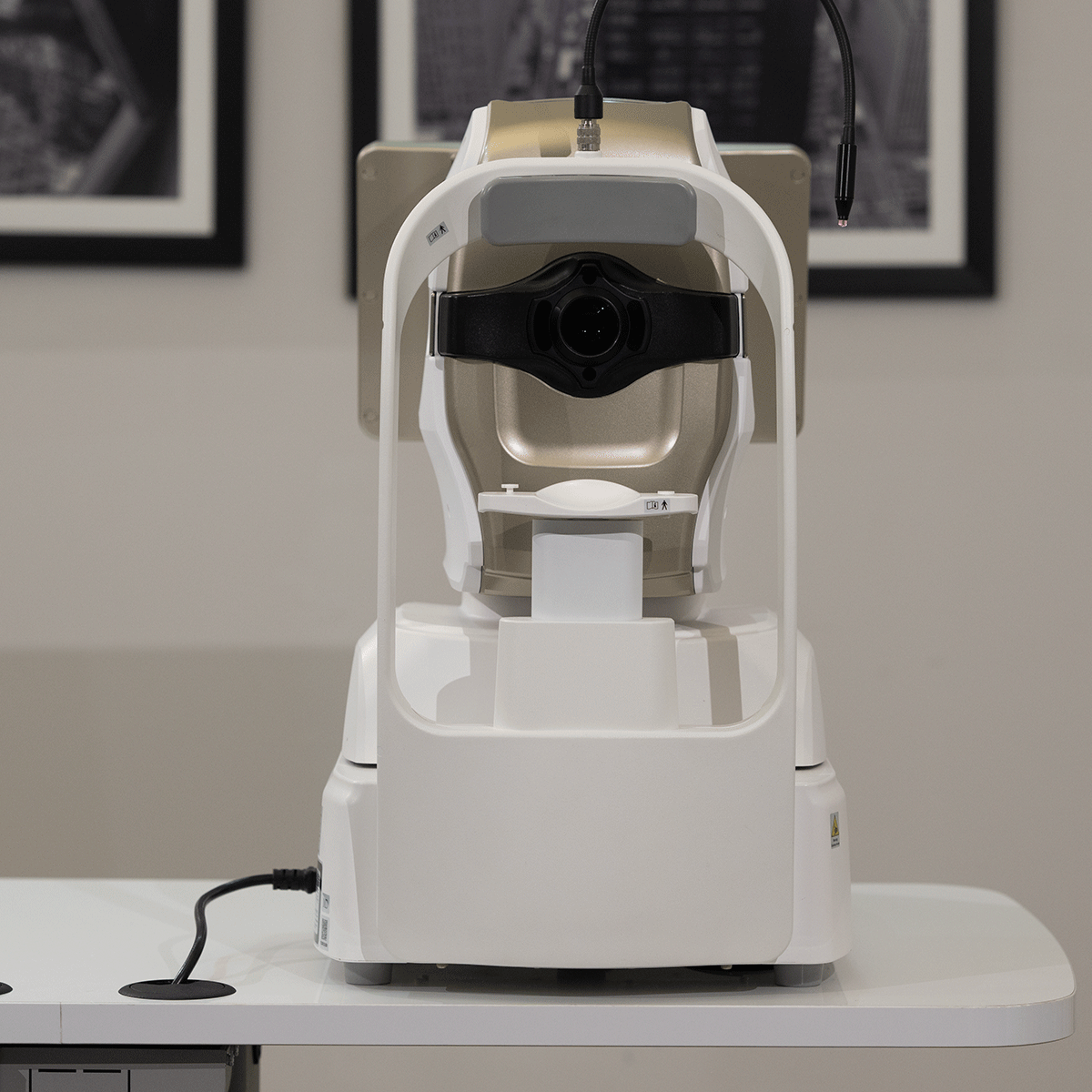
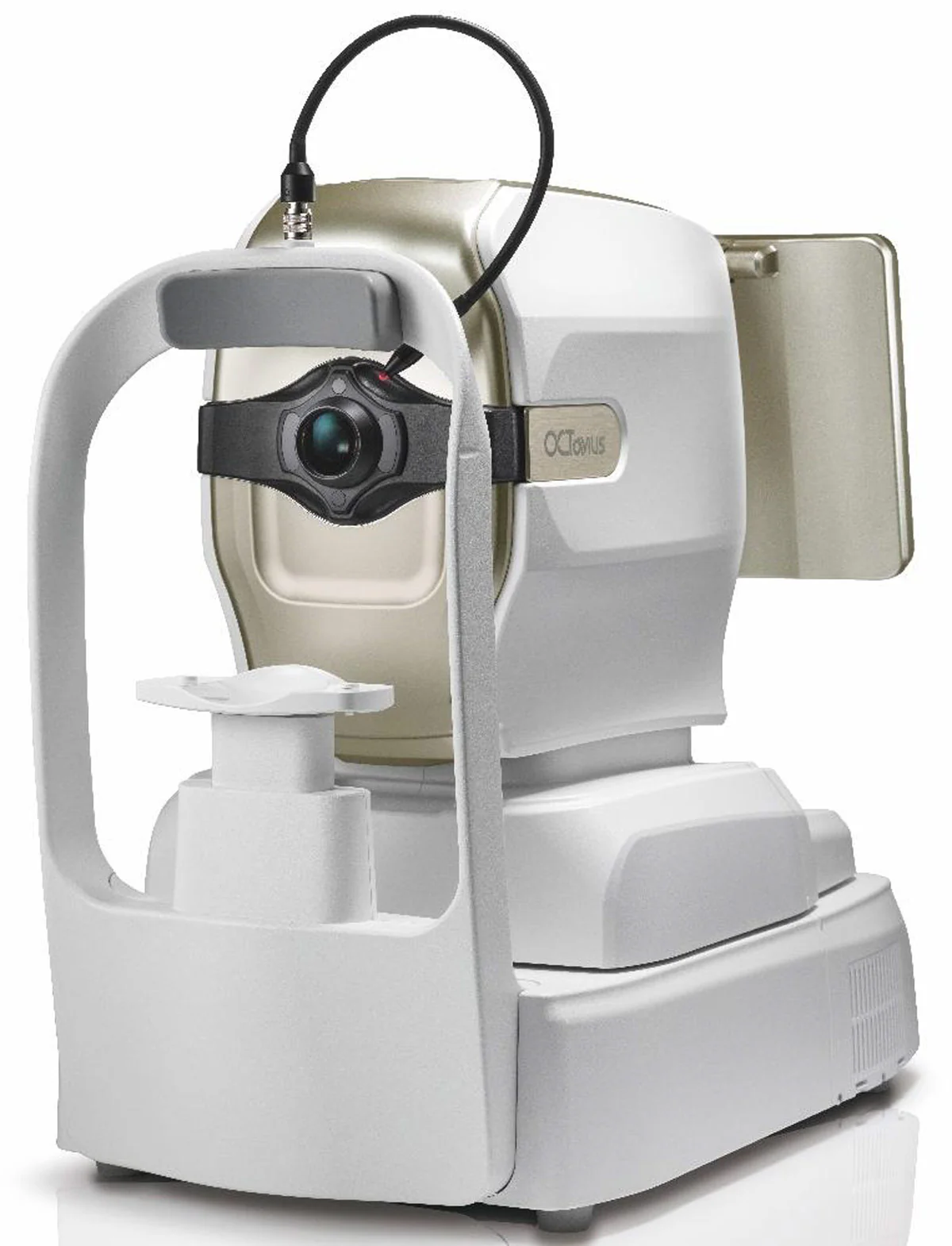
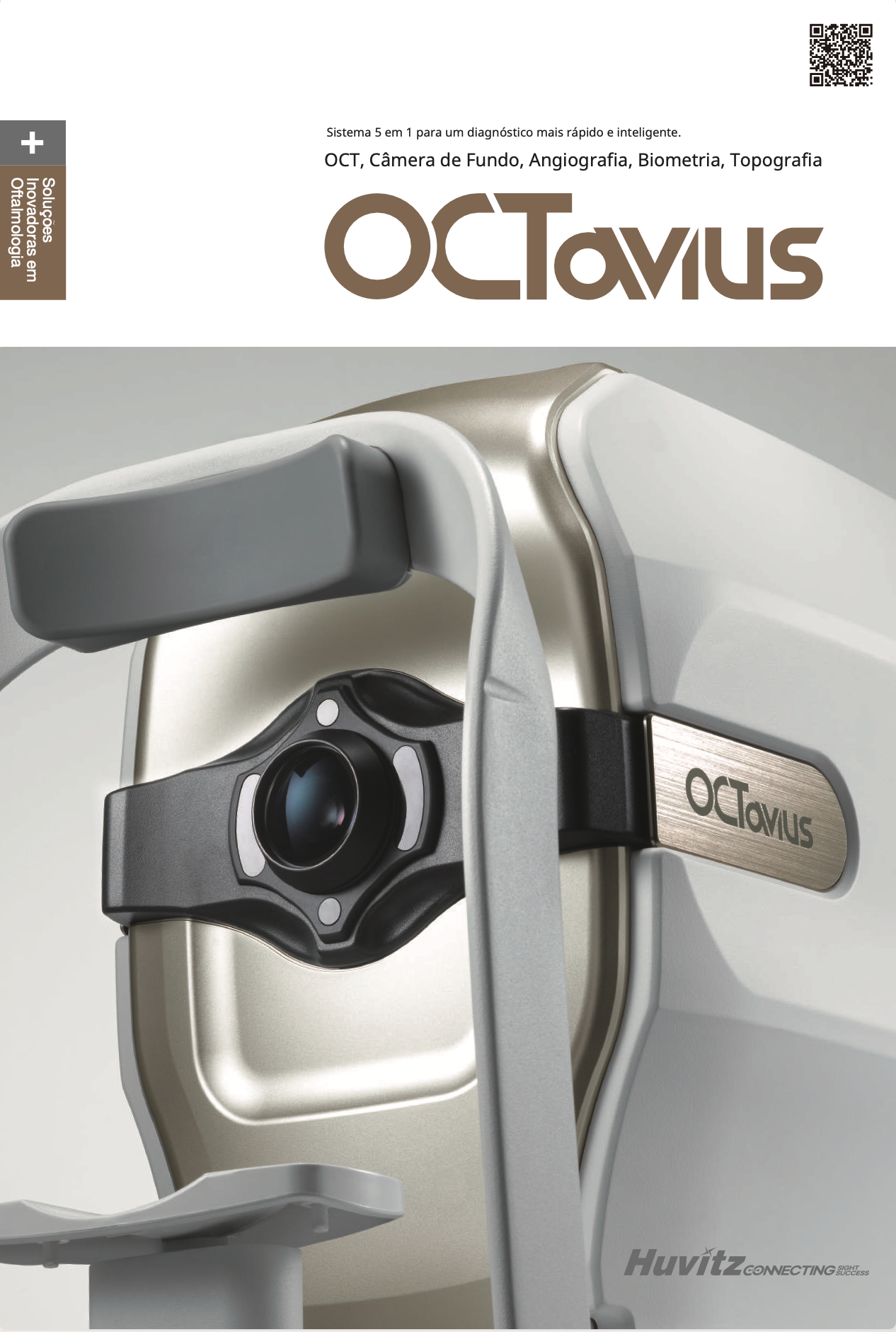
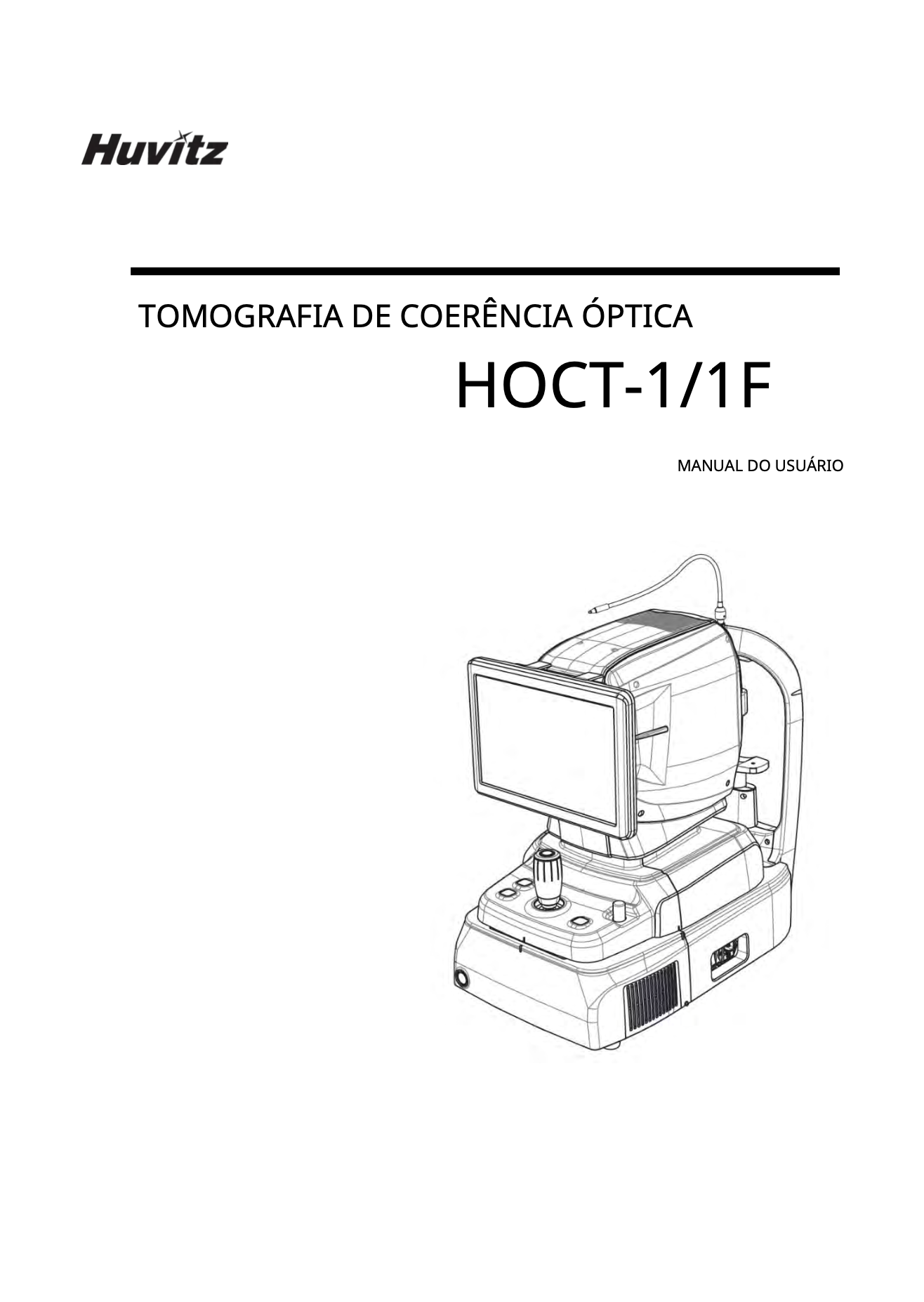
OCT
-
Principle
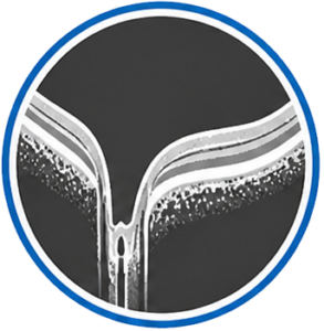
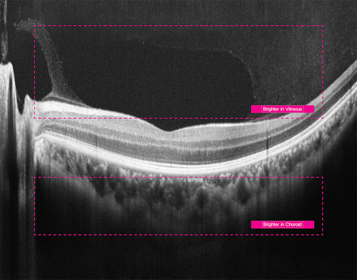
Spectral domain OCT
-
Light source
840 nm
-
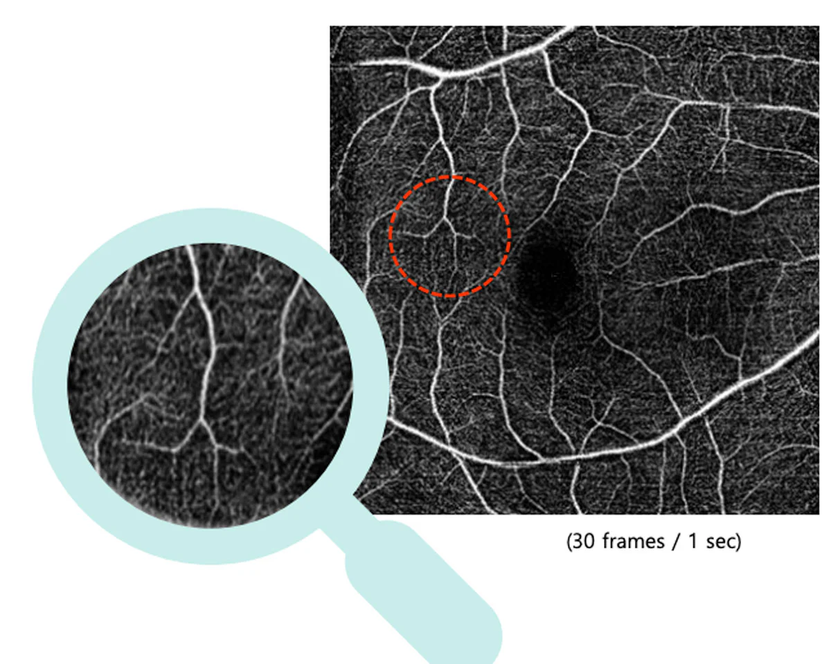
Scanning speed
Max. 80,000 A-scans/sec.
-
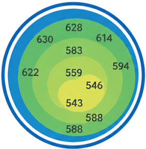
Resolution in the fabric
20 μm (lateral), 7 μm (z-axis) with index 1.36
-
Scan range
X : 6~12mm, Y : 6~9mm, Z : 2.34mm
-
Display resolution
X: 5.85μm, Y: 23.40μm, Z: 3.05μm
-
Minimum pupil diameter
2.5 mm
-
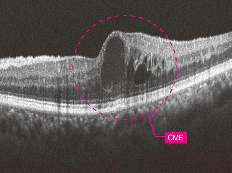
Scanning patterns
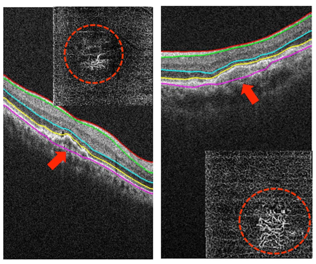
Macula: Macular line, Macular cross, Macular radial, 3D macula, Macular raster, Angio (optional)
Disc: Disc circle, Disc radial, 3D disc, Disc raster, Angio (optional)
-
Optical power in the cornea
≤ 1.3 mW
-
3D image acquisition time
1.0 s (Normal mode, A512xB96)
-
Depth accuracy (measurement in glass to 1 mm)
±3%
-
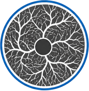
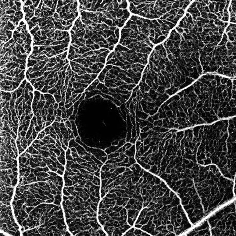
OCT Angiography – Option (HOCT-1/1F)
-
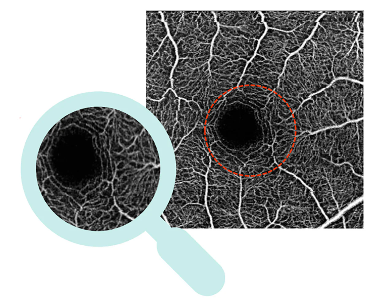
Angiography band
3-9 mm
-
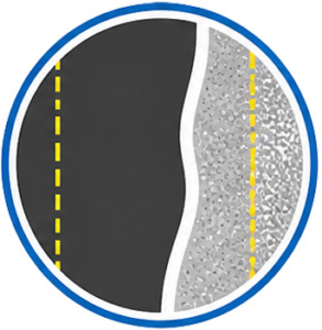
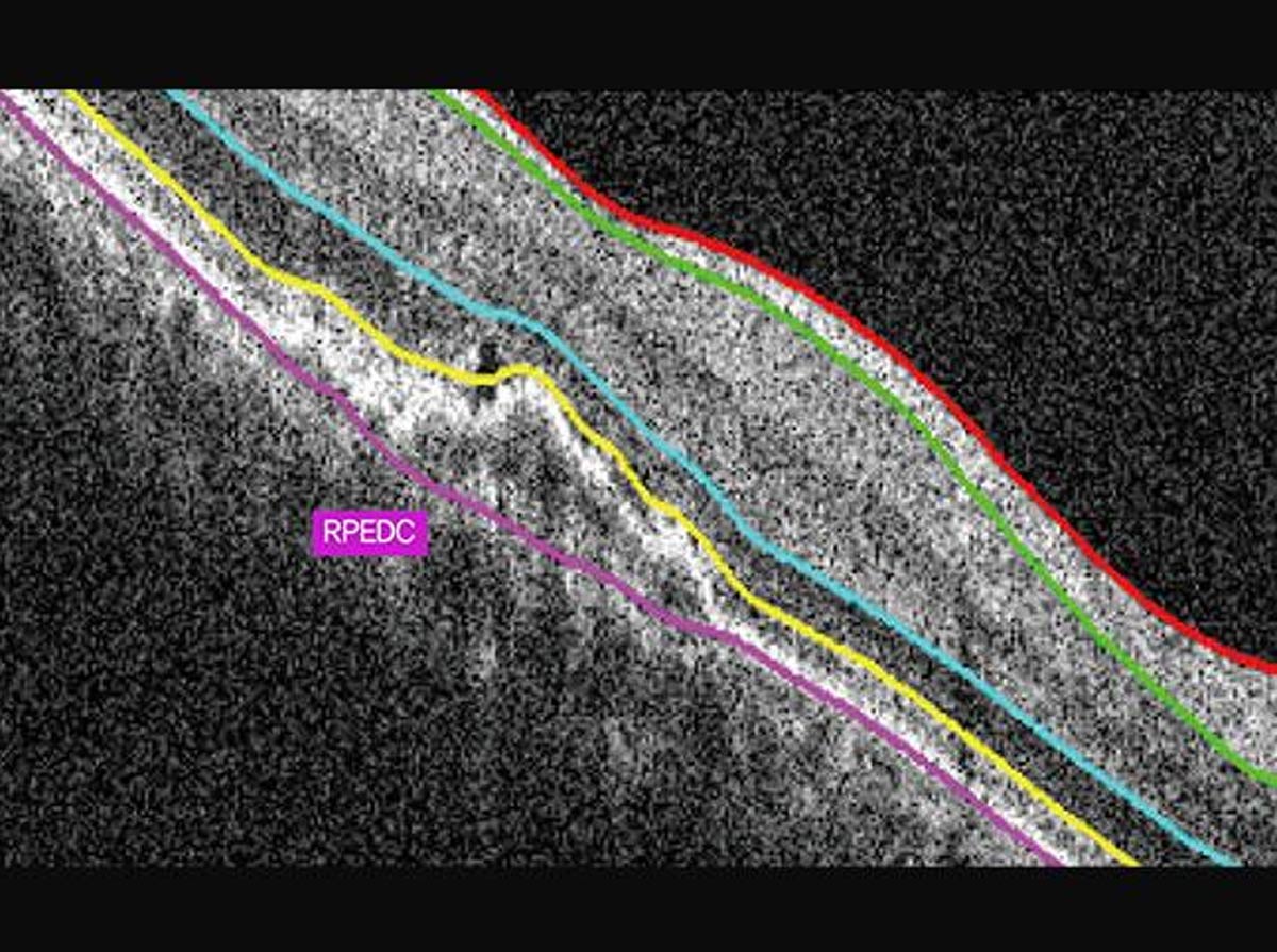
Angiography map
Superficial, Deep, External, Choriocapillary, Retina, Customized, Enface, Thickness Map, Depth-coded Map
-
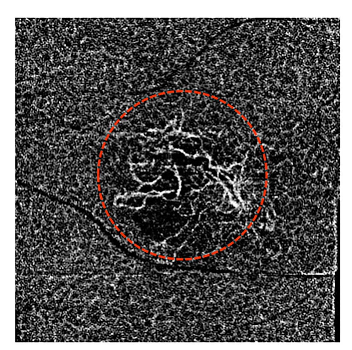
Angiography analysis
FAZ, Vessel density
-
Common specifications
-
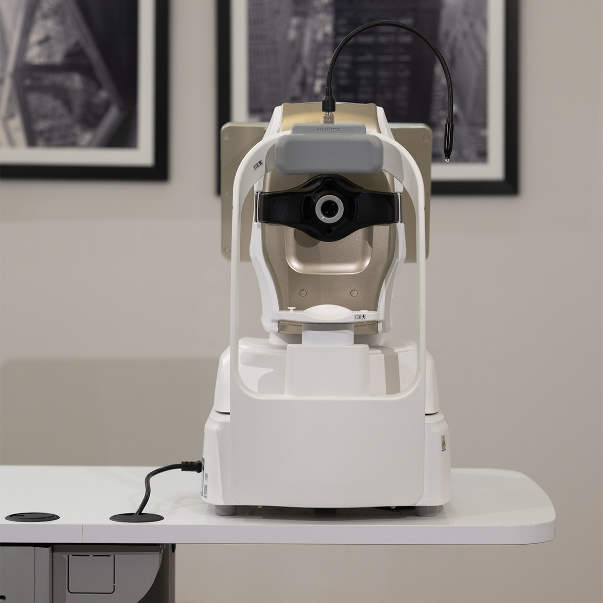
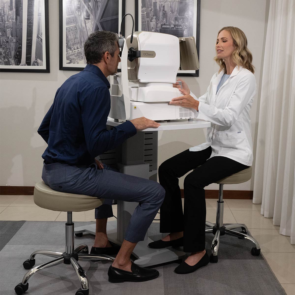
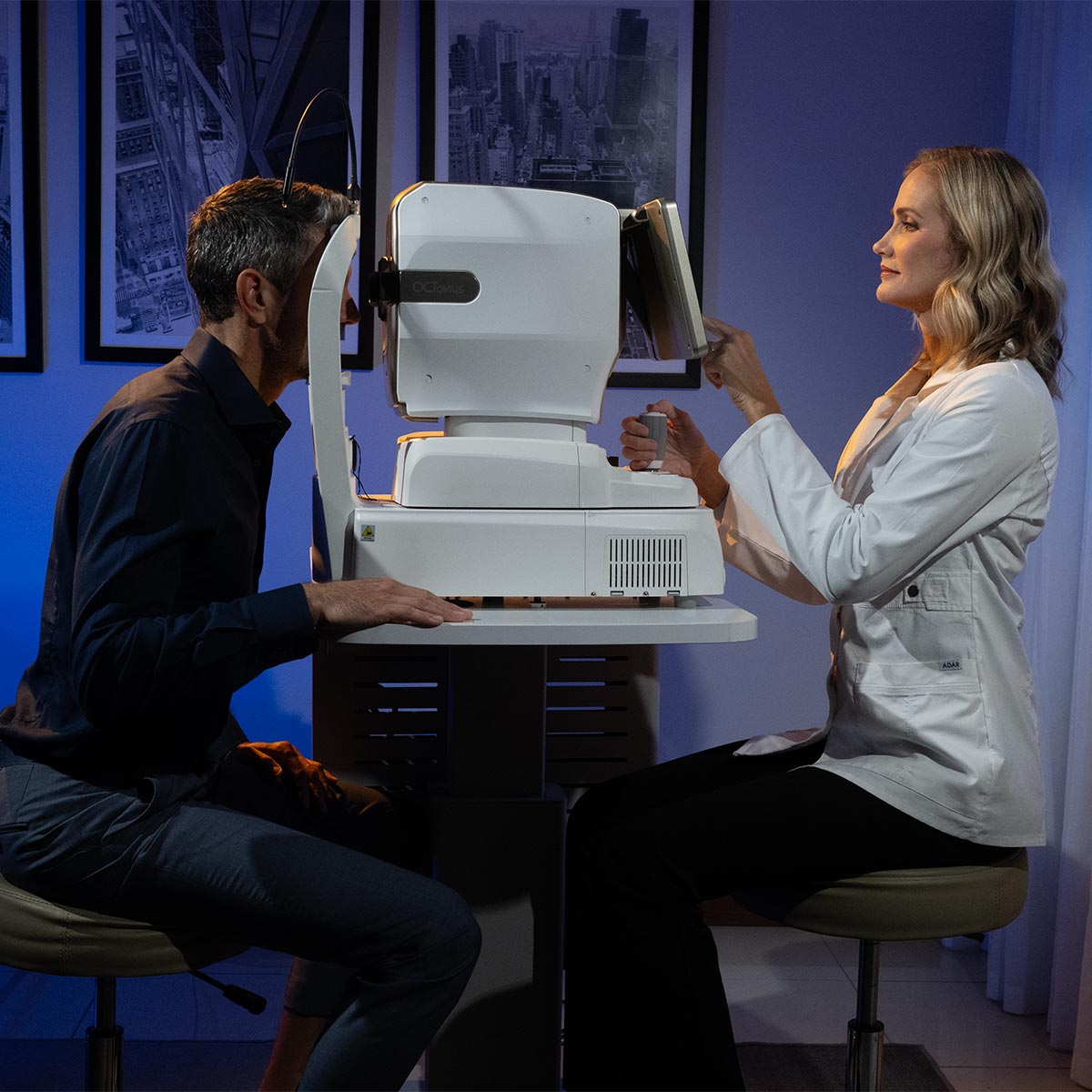
Working distance
33 mm
-
33 mm
12.1-inch, 1280 x 800 pixel, color touchscreen LCD panel.
-
Dioptric compensation of the patient's eye
-33D~+33D total, -13D~+13D without corrective lenses, +7D~+33D with positive lenses, -33D~-7D with negative lenses
-
Target for attachment
LCD (internal), white LED (external)
-
Horizontal movement
70 mm (front and back), 100 mm (left and right)
-
Vertical movement
30 mm
-
Chin rest movement
62 mm (up and down), motorized
-
Self-tracking
30 mm (up and down), 10 mm (right and left), 10 mm (front and back)
-
30 mm (up and down), 10 mm (right and left), 10 mm (front and back)
AC 100 - 240 V, 50/60 Hz, 1.6 - 0.7 A
-
PRAÇA
Integrated computer
-
LCD tilt angle
70˚
-
Dimensions / Weight
330 (W) x 542 (D) x 521 (H) mm / 30 kg
-
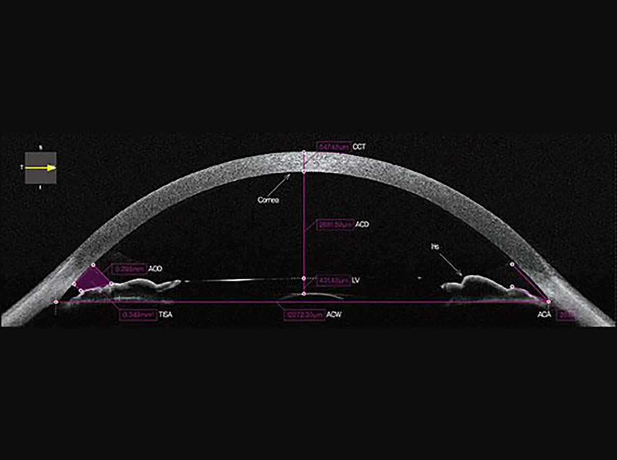
Anterior segment adapter (optional)
-
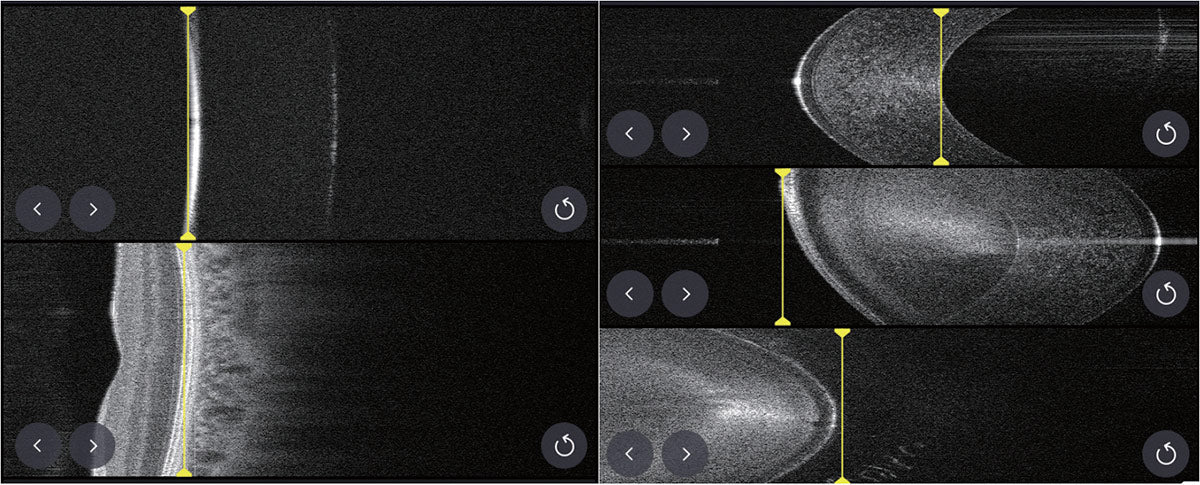
Working distance
15 mm (from the anterior segment adapter to the eyeball)
-
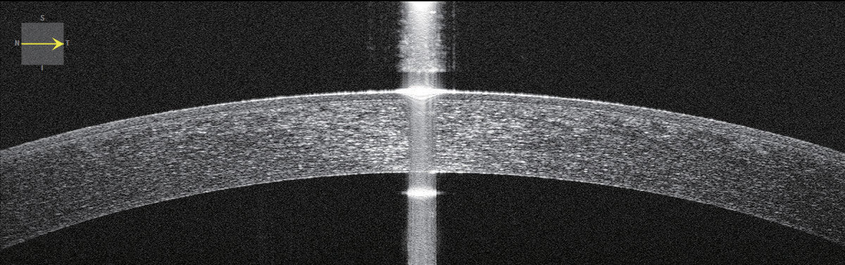
Scan range
6 ~ 9 mm (width), 2.3 mm (depth)
-
Scan pattern
ACA line, anterior radial
-
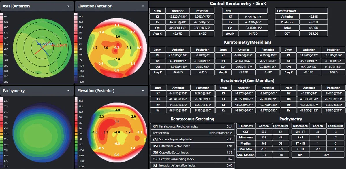
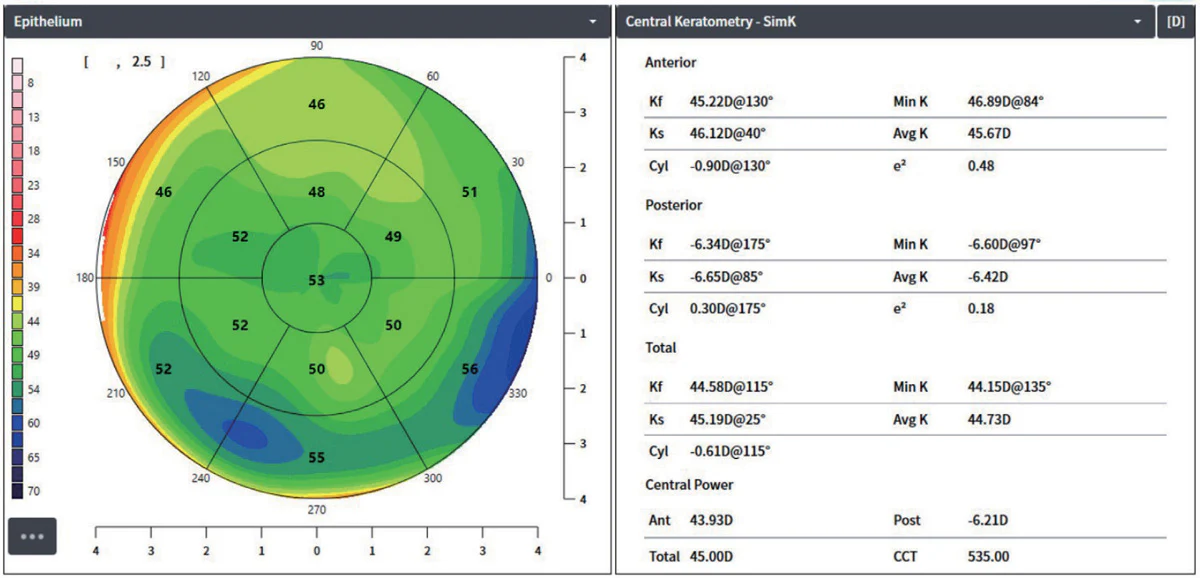
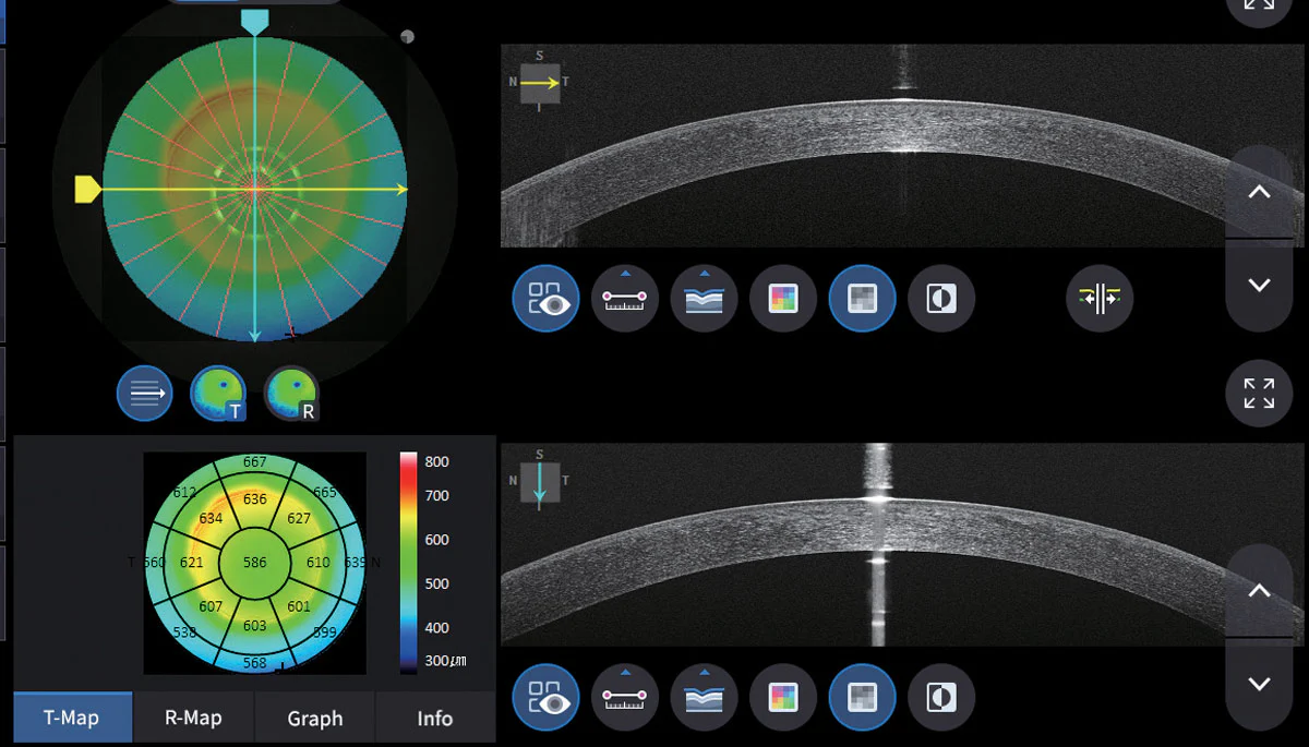
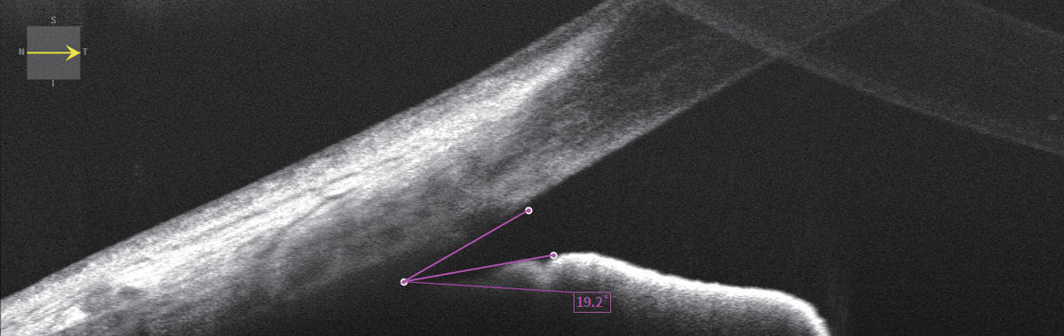
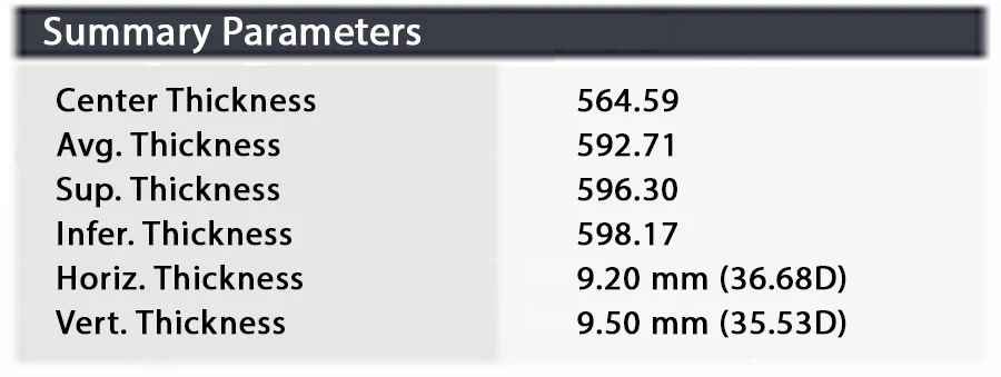
Metric
Corneal layers, Thickness map, Thickness, Angle
Wide anterior segment adapter (optional)
-
Working distance
15 mm
-
Scan range
16 mm (width), 2.3 mm (depth)
-
Scan pattern
ACA line, Front radial, Complete
-
Metric
Dimension, Angle
-
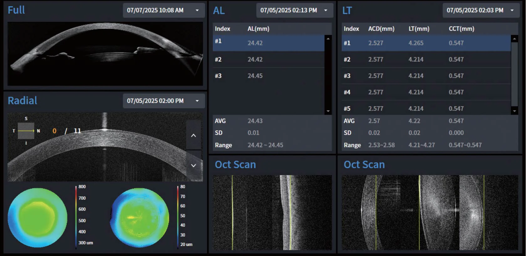
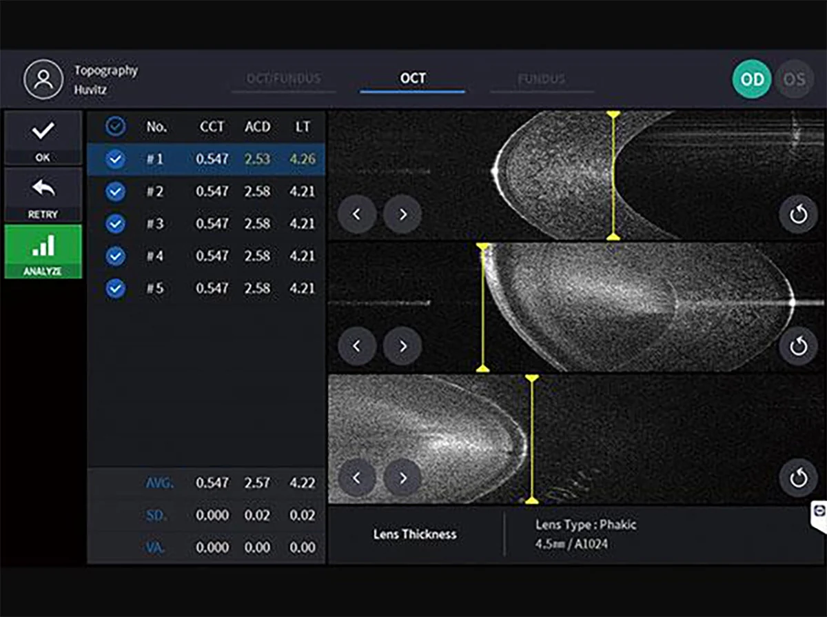
Biometrics (optional)
-
Metric
AL, CCT, ACD, LT
-
Topography (optional)
-
Compatible maps
Axial map, Tangential map, Keratoconus detection
-
HIIS-1 (optional)
-
Function
Web-based, accessible to multiple users, progression analysis, comparative analysis, 3D analysis.